DNA
Molecules of life
Deoxyribonucleic acid (DNA) forms the genetic basis of every organism on this planet. Whether unicellular organism, plant or animal – as the carrier of genetic information, this macromolecule not only determines appearance, but also life expectancy, state of health and, of course, gender. The unbelievable diversity of our earth as we know it today has developed from only four individual building blocks, the nucleotides, through billions of years of evolution from one single cell, e.g. the last universal cellular ancestor (LUCA).
Adenine
thymine
guanine
cytosine
nucleobases
nucleotide
Chemical basis
The basic structure of DNA consists of the four bases adenine, thymine, guanine and cytosine. Adenine and guanine belong to the group of purines. Thymine and cytosine are pyrimidines. Each of these nucleobases are connected to a C5 sugar through the C1 atom via an N-glycosidic bond. At the exposed C5, this sugar attaches to a phosphate residue by forming an ester bond. When pyrophosphate (two phosphate residues) is cleaved off, thousands of nucleotides can line up via the sugar backbone to form a single DNA strand containing specific information.

G (5')
C (3')
T
A
T
G
C
A
A
C
G
T
C
G
T (3')
A (5')
biological design
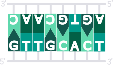
Physiologically, DNA is found in the form of a right-handed double strand. In each case, complementary base pairs are coupled. Adenine (A) is stabilized by two hydrogen bonds with thymine (T). Guanine (G) forms three hydrogen bonds to cytosine (C). As E. Chargaff found out in the middle of the 20th century, in each cell the amount of Adenine is equal to Thymine and C = G (parity rule). Furthermore, both strands run antiparallel from the sugar backbone. In order to save space, the DNA twists around its own axis, creating the typical double-helix form.
key figures
One helix winding has a length of 3.4 nm or 34 Ångström. With a twist angle of 36° between the individual planes, exactly 10 base pairs form a complete turn. The sugar-phoshpate-backbone is located outside and is a sufficient condition that each wound consists of a major groove with a height of 2.2 nm and a minor groove with a height of 1.2 nm. This is necessary to enable different proteins to bind to DNA strands. For example, transcription factors to transcribe a specific gene, or histone proteins to further package the double strand.
A-DNA
B-DNA
Z-DNA
Physiological variations
At a hydration rate of over 95%, including the environmental conditions of human cells, the DNA is present in the B form. The complementary base pairs face each other and twist around their own axis. If the degree of hydration drops, the conformation changes to the A-form. Here the sugar changes from 2′-endo to 3′-endo conformation. The base pairs are oriented around the axis of rotation. The A-DNA is 30% shorter and wider than B-DNA. Z-DNA is metastable and can be observed at high salt concentrations. It is left-handed and its physiological occurence has not been clarified.
imprint
Research, design and publication by
Peter Schönhammer,
M. Sc. Biochemistry, University of Bayreuth, 2019.
License: CC BY-NC-SA 4.0.
References:
Knippers, Rolf (2006): Molekulare Genetik. 9., komplett überarb. Aufl. Stuttgart: Thieme. ISBN: 9783134770094
Berg, Jeremy M., Tymoczko, John L., Stryer, Lubert: Stryer Biochemie, Springer Berlin Heidelberg, January 2013. ISBN: 978-3-82-742988-9